Nye publikasjoner
Miniatyrsonde for optisk koherens-tomografi tar bilder inne i hjernearteriene
Sist anmeldt: 02.07.2025

Alt iLive-innhold blir gjennomgått med medisin eller faktisk kontrollert for å sikre så mye faktuell nøyaktighet som mulig.
Vi har strenge retningslinjer for innkjøp og kun kobling til anerkjente medieområder, akademiske forskningsinstitusjoner og, når det er mulig, medisinsk peer-evaluerte studier. Merk at tallene i parenteser ([1], [2], etc.) er klikkbare koblinger til disse studiene.
Hvis du føler at noe av innholdet vårt er unøyaktig, utdatert eller ellers tvilsomt, velg det og trykk Ctrl + Enter.

Et internasjonalt team av mikroteknologer, medisinske teknologer og nevrokirurger har designet, bygget og testet en ny type sonde som kan brukes til å ta bilder fra innsiden av hjernens arterier.
I artikkelen sin publisert i tidsskriftet Science Translational Medicine beskriver gruppen hvordan sonden ble designet og bygget, samt hvordan den presterte i innledende tester.
Når pasienter har medisinske problemer i hjernen, som blodpropper, aneurismer eller forkalkede arterier, er verktøyene som er tilgjengelige for leger for å diagnostisere dem begrenset til bildebehandlingsteknologier som tar bilder av vener og arterier fra utsiden av hjernen. Disse bildene brukes deretter som kart for å lede kateterlignende enheter gjennom venene og arteriene inn i deler av hjernen for å utføre reparasjoner.

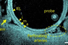
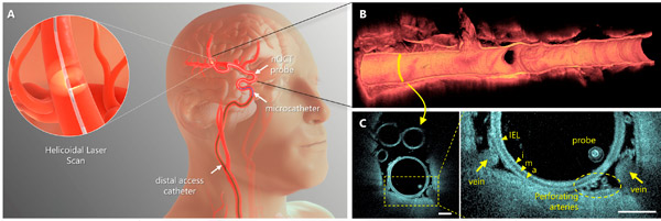
Intravaskulær avbildning ved bruk av nevrooptisk koherenstomografi (nOCT). nOCT-sonden er kompatibel med standard nevrovaskulære mikrokatetre og integreres med den prosedyremessige arbeidsflyten som brukes i klinisk praksis. nOCT fanger opp høyoppløselige 3D-optiske data, og gir volummikroskopi av kronglete hjernearterier, omkringliggende strukturer og terapeutiske apparater. Kilde: Science Translational Medicine (2024). DOI: 10.1126/scitranslmed.adl4497
Problemet med denne tilnærmingen er at bildene som brukes ikke alltid er klare eller nøyaktige. De lar heller ikke kirurgen se hva som skjer inne i venen eller arterien mens den repareres, noe som resulterer i at prosedyrene utføres nesten blindt.
I denne nye studien laget teamet en kamerasonde som er liten nok til å passe inn i et kateter, slik at den kan ta bilder i nær sanntid fra innsiden av hjernens vener og arterier.
Den nye sonden er basert på optisk koherenstomografi, en type bildebehandlingsteknologi som brukes av øye- og hjertekirurger for å behandle pasienter. Den genererer bilder ved å behandle tilbakespredningen av nær-infrarødt lys. Frem til nå har slike enheter vært for store og stive til å kunne brukes inne i hjernen.
For å løse dette problemet byttet forskerteamet ut komponentene med mindre deler, som en fiberoptisk kabel så tynn som et menneskehår. De brukte også en modifisert type glass for å lage den distale linsen, som utgjør hodet på proben og lar den bøye seg.
Den resulterende sonden er stort sett hul og ormformet. Den roterer også 250 ganger per sekund, noe som gjør at den beveger seg enkelt gjennom venene og arteriene. Kameraet tar bilder med en hastighet proporsjonal med behovet. Hele sonden passer lett inn i kateteret, noe som gjør det enkelt å plassere og flytte rundt i arteriene og venene i hjernen, samt å fjerne den.
Etter dyreforsøk ble proben flyttet til kliniske studier på to steder, ett i Canada og ett i Argentina. Til dags dato har 32 pasienter blitt behandlet med den nye proben. Teamet rapporterer at proben så langt har vist seg trygg, godt tolerert og vellykket i alle tilfeller. De konkluderer med at den nye proben er klar for generell bruk.
