Nye publikasjoner
Kunstig intelligens-verktøy avslører kjønnsforskjeller i hjernens struktur
Sist anmeldt: 02.07.2025

Alt iLive-innhold blir gjennomgått med medisin eller faktisk kontrollert for å sikre så mye faktuell nøyaktighet som mulig.
Vi har strenge retningslinjer for innkjøp og kun kobling til anerkjente medieområder, akademiske forskningsinstitusjoner og, når det er mulig, medisinsk peer-evaluerte studier. Merk at tallene i parenteser ([1], [2], etc.) er klikkbare koblinger til disse studiene.
Hvis du føler at noe av innholdet vårt er unøyaktig, utdatert eller ellers tvilsomt, velg det og trykk Ctrl + Enter.

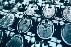
Kunstig intelligens (KI) dataprogrammer som behandler MR-skanninger avslører forskjeller i organiseringen av hjernen til menn og kvinner på cellenivå, viser en ny studie. Disse forskjellene ble funnet i hvit substans, vevet som hovedsakelig finnes i det indre laget av den menneskelige hjernen som legger til rette for kommunikasjon mellom regioner.
Menn og kvinner er kjent for å lide ulikt av multippel sklerose, autismespekterforstyrrelse, migrene og andre hjerneproblemer, og for å vise forskjellige symptomer. En detaljert forståelse av hvordan biologisk kjønn påvirker hjernen blir sett på som en måte å forbedre diagnostiske verktøy og behandlinger på. Selv om hjernens størrelse, form og vekt har blitt studert, har forskere bare en delvis forståelse av dens struktur på cellenivå.
En ny studie ledet av forskere ved NYU Langone Health brukte en AI-teknikk kalt maskinlæring til å analysere tusenvis av MR-hjerneskanninger fra 471 menn og 560 kvinner. Resultatene viste at dataprogrammene nøyaktig kunne skille mellom mannlige og kvinnelige hjerner, og identifisere strukturelle og komplekse mønstre som var usynlige for det menneskelige øyet.
Resultatene ble bekreftet av tre forskjellige AI-modeller designet for å bestemme biologisk kjønn, ved å bruke deres relative styrker enten ved å fokusere på små flekker av hvit substans eller analysere forbindelser på tvers av store områder av hjernen.
«Funnene våre gir et klarere bilde av strukturen til den levende menneskehjernen, noe som kan gi ny innsikt i hvordan mange psykiatriske og nevrologiske lidelser utvikler seg og hvorfor de kan manifestere seg forskjellig hos menn og kvinner», sa hovedforfatter av studien og nevroradiolog Yvonne Lui, MD.
Lui, professor og nestleder for forskning ved radiologiavdelingen ved NYU Grossman School of Medicine, bemerker at tidligere studier av hjernens mikrostruktur i stor grad har vært avhengige av dyremodeller og menneskelige vevsprøver. I tillegg har gyldigheten av noen av disse tidligere funnene blitt stilt spørsmål ved ved bruk av statistiske analyser av «håndtegnede» interesseområder, som krevde at forskere tok mange subjektive beslutninger om formen, størrelsen og plasseringen av regionene de valgte. Slike valg kan potensielt skjeve resultatene, sier Lui.
Funnene i den nye studien unngikk dette problemet ved å bruke maskinlæring til å analysere hele grupper av bilder uten å fortelle datamaskinen at den skulle se på et bestemt sted, noe som bidro til å eliminere menneskelige skjevheter, bemerker forfatterne.
For studien startet teamet med å mate AI-programmene med eksisterende data, for eksempel MR-hjerneskanninger av friske menn og kvinner, sammen med det biologiske kjønnet til hver skanning. Fordi disse modellene var designet for å bruke sofistikerte statistiske og matematiske metoder for å bli «smartere» over tid etter hvert som de samlet inn data, «lærte» de til slutt å skjelne biologisk kjønn på egenhånd. Viktigere er det at programmene var begrenset fra å bruke generell hjernestørrelse og -form for sine bestemmelser, sier Lui.
Ifølge resultatene identifiserte alle modellene kjønnet på skanningene korrekt i 92 % til 98 % av tilfellene. Flere funksjoner hjalp maskinene med å trekke konklusjonene sine, inkludert hvor lett og i hvilken retning vann kunne bevege seg gjennom hjernevevet.
«Disse funnene fremhever viktigheten av mangfold når man studerer sykdommer som stammer fra den menneskelige hjerne», sa studiens medforfatter Junbo Chen, MS, en doktorgradsstudent ved NYU Tandon School of Engineering.
«Hvis menn brukes som standardmodell for ulike lidelser, slik det historisk sett har vært tilfelle, kan forskere gå glipp av kritisk innsikt», la studiens medforfatter Vara Lakshmi Bayanagari, MS, en forskerstudent ved NYU Tandon School of Engineering, til.
Bayanagari advarer om at selv om AI-verktøyene kunne rapportere forskjeller i hjernecelleorganisering, kunne de ikke identifisere hvilket kjønn som var mer utsatt for hvilke egenskaper. Hun legger til at studien klassifiserte kjønn basert på genetisk informasjon og kun inkluderte MR-skanninger av cisgender menn og kvinner.
Teamet planlegger å studere utviklingen av kjønnsforskjeller i hjernestrukturen videre over tid for å bedre forstå rollen til miljømessige, hormonelle og sosiale faktorer i disse endringene, sier forfatterne.
Arbeidet ble publisert i tidsskriftet Scientific Reports.
